Resolving brain architecture with comprehensive spatial gene expression
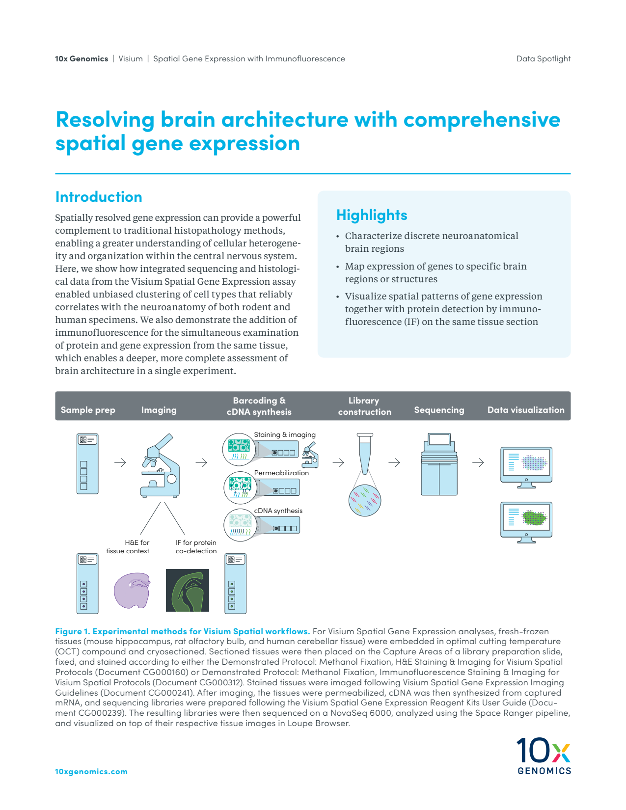
Spatially resolved gene expression can provide a powerful complement to traditional histopathology methods, enabling a greater understanding of cellular heterogeneity and organization within the central nervous system. Here, we show how integrated sequencing and histological data from the Visium Spatial Gene Expression assay enabled unbiased clustering of cell types that reliably correlates with the neuroanatomy of both rodent and human specimens. We also demonstrate the addition of immunofluorescence for the simultaneous examination of protein and gene expression from the same tissue, which enables a deeper, more complete assessment of brain architecture in a single experiment.
Spatially resolved gene expression can provide a powerful complement to traditional histopathology methods, enabling a greater understanding of cellular heterogeneity and organization within the central nervous system. Here, we show how integrated sequencing and histological data from the Visium Spatial Gene Expression assay enabled unbiased clustering of cell types that reliably correlates with the neuroanatomy of both rodent and human specimens. We also demonstrate the addition of immunofluorescence for the simultaneous examination of protein and gene expression from the same tissue, which enables a deeper, more complete assessment of brain architecture in a single experiment.