Uncover the "Where" for every "What"
The relationship between cells and their relative locations within tissue is critical to understanding normal development and disease pathology. Spatial transcriptomics is a groundbreaking molecular profiling method that allows scientists to measure all the gene activity in a tissue sample and map where the activity is occurring. Already this technology is leading to new discoveries that are proving instrumental in helping scientists gain a better understanding of biological processes and disease.
Fuel your spatial discoveries with spatial capture technology
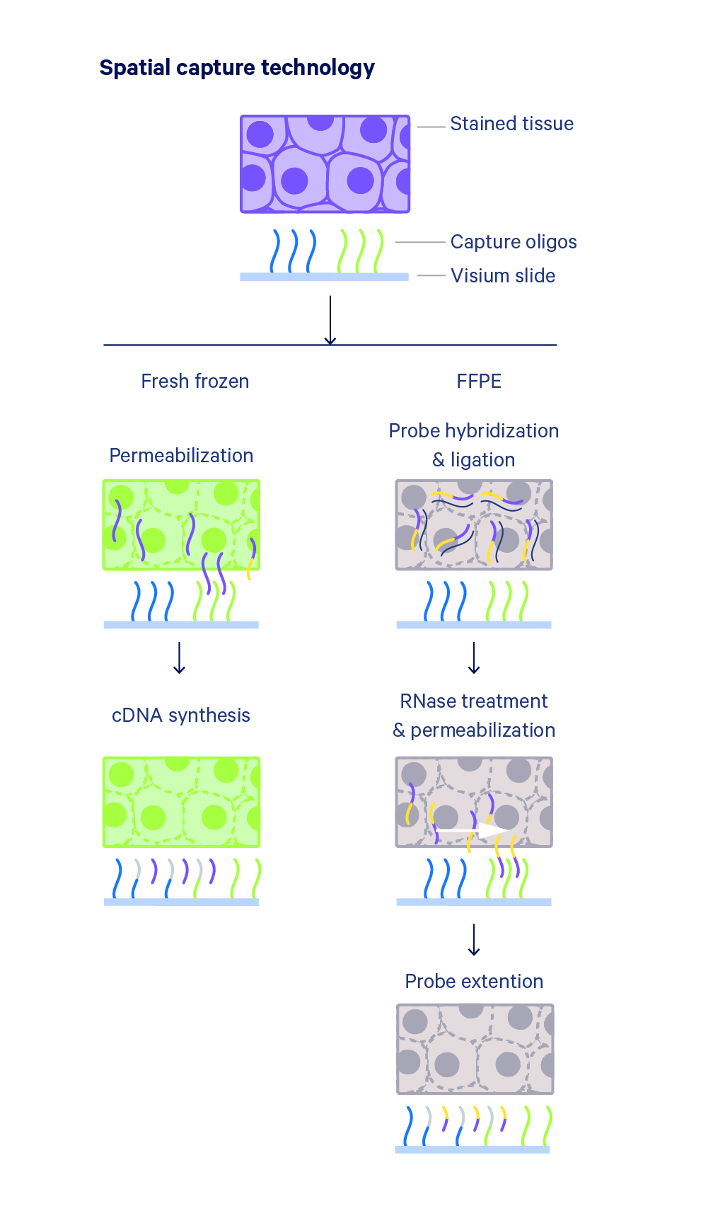
Spatial capture technology powers the Visium Spatial platform through the use of spatially barcoded mRNA-binding oligonucleotides. There are two methods for how mRNA molecules get a spatial barcode.
In fresh frozen tissues, the tissue is fixed and permeabilized to release RNA which binds to adjacent capture probes, allowing for the capture of gene expression information. cDNA is then synthesized from captured RNA and sequencing libraries prepared.
In FFPE tissues, the tissue is permeabilized to release ligated probe pairs that bind to adjacent capture probes on the slide, allowing for the capture of gene expression information. Pairs of probes specific to each gene in the protein-coding transcriptome are hybridized to their gene target and then ligated to one another. The probe pairs are extended to incorporate complements of the spatial barcodes and sequencing libraries prepared.

Examine gene expression in the context of the tissue microenvironment
Innovation in spatial transcriptomics methodologies is enabling scientists to get a holistic understanding of cells in their morphological context. In this presentation, you will hear first-hand from 10x Genomics scientists about groundbreaking improvements to the technology and exciting applications showcased by users.
Gain a holistic understanding of gene and protein expression in the tissue microenvironment
Versican promotes T helper 17 cytotoxic inflammation and impedes oligodendrocyte precursor cell remyelination
Versican promotes T helper 17 cytotoxic inflammation and impedes oligodendrocyte precursor cell remyelination
Ghorbani S, et al. Nature Communications 2022.
Ghorbani S, et al. Nature Communications 2022.
Single-cell and spatial analysis reveal interaction of FAP+ fibroblasts and SPP1+ macrophages in colorectal cancer
Single-cell and spatial analysis reveal interaction of FAP+ fibroblasts and SPP1+ macrophages in colorectal cancer
Qi J, et al. Nature Communications 2022.
Qi J, et al. Nature Communications 2022.
Spatial transcriptomics of dorsal root ganglia identifies molecular signatures of human nociceptors
Spatial transcriptomics of dorsal root ganglia identifies molecular signatures of human nociceptors
Tavares-Ferreira D, et al. Science Translational Medicine 2022.
Tavares-Ferreira D, et al. Science Translational Medicine 2022.
Explore data visualization
Visualization Controls
Use the sliders under the tissue image to adjust how you visualize and combine the tissue image and the gene expression data. Colors represent clusters identified by differentially expressed genes.
Gene Identification
By placing the pointer above a gene name within the table, spots in the tissue image will be colored based on the expression of that gene. Alternatively, by placing the pointer above a value within the table, you can observe the expression of a specific gene with the spots from an individual cluster highlighted.
Beyond gene expression
Layer transcriptome and proteome data onto your stained images to understand tissue microenvironments like never before. Reveal the spatial organization of newly discovered cell types, states, and biomarkers.Turnkey solution
Compatible with diverse sample types across preparation (FFPE and fresh frozen) and species (human, mouse, rat, and more), easily incorporate into your research with all the reagents kitted and ready to use and no specialized instrumentation required.Easily adoptable workflow
Integrate easily with your current laboratory methods and tools for tissue analysis. Combine with immunofluorescence staining and imaging to gain multiomic characterization in the spatial context.Streamlined data analysis
Analyze and understand gene and protein expression heterogeneity with Space Ranger analysis software to process your data, and interactively explore the results with Loupe Browser visualization software.