“What limits our progress in biological, biomedical sciences? It's the technologies. Science takes a big leap forward every time there's a new technology.”
Building subcellular spatial maps with Xenium In Situ
The Xenium platform achieves highly specific and sensitive detection of RNA targets using a probe-based strategy. The probes are designed using a proprietary 10x Genomics algorithm that ensures no probe crosstalk.
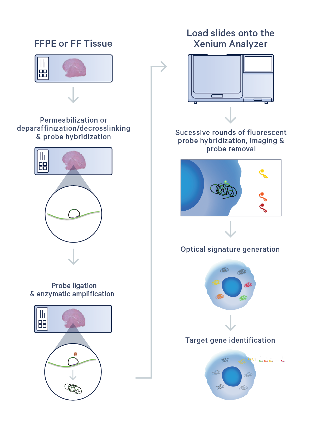
Sample processing starts with sectioning tissues onto a Xenium slide. Sections are then treated to access the RNA for labeling with circularizable DNA probes. Ligation of the probes then generates a circular DNA probe which is enzymatically amplified and bound with fluorescent oligos, creating a bright, easy-to-image signal that has a high signal-to-noise ratio. Up to two slides are then placed in the Xenium Analyzer where the samples undergo successive rounds of fluorescent probe hybridization, imaging, and removal. An optical signature specific to each gene is generated, enabling identification of the target gene. Finally, a spatial map of the transcripts is built across the entire tissue section.
Explore the data
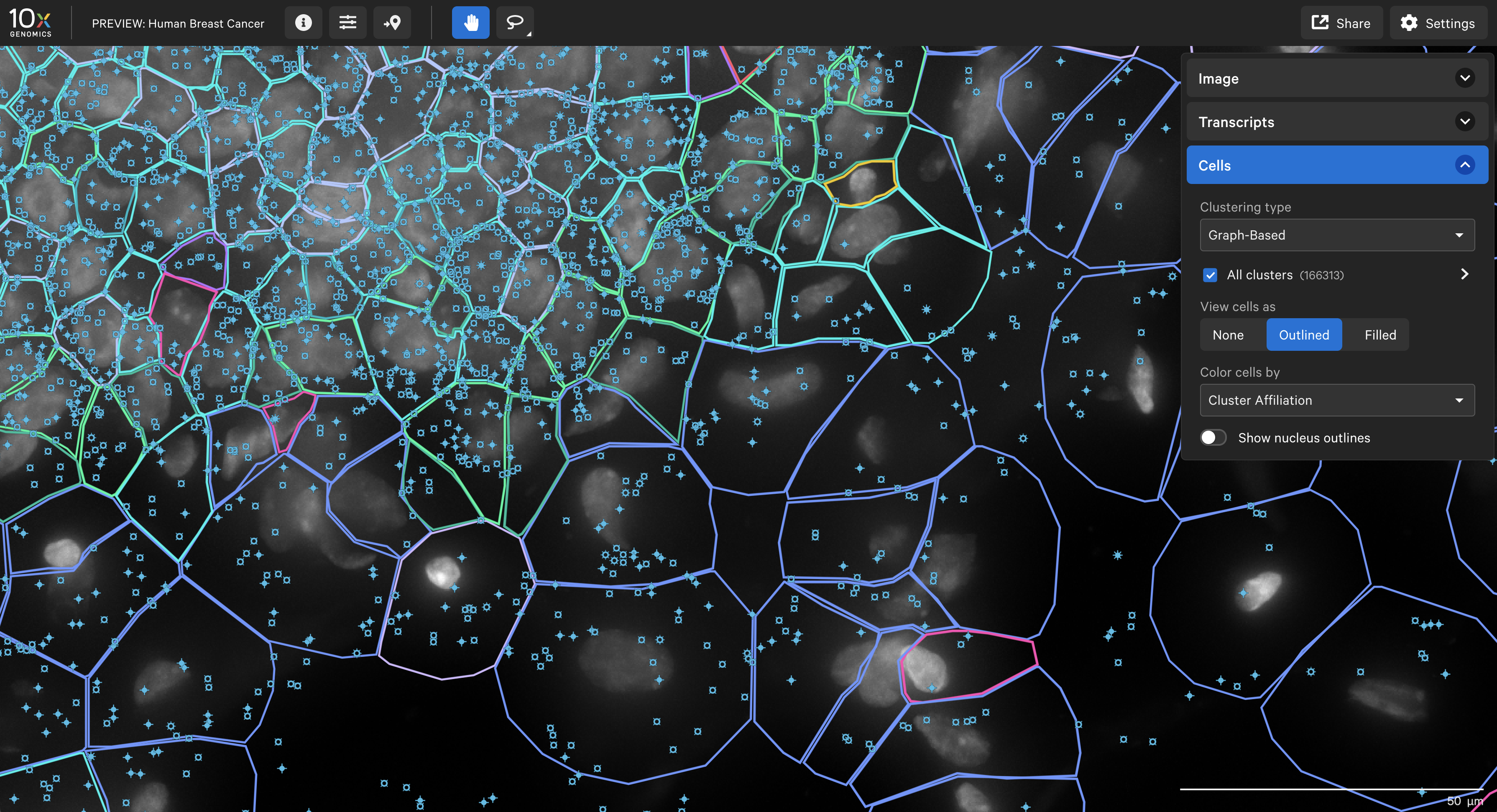
- Use Xenium Explorer Demo, a preview of the upcoming desktop app, to explore a Xenium In Situ dataset from a human breast cancer sample using our 280-gene Xenium Human Breast Gene Expression Panel with 33 additional custom genes.
The end-to-end Xenium platform
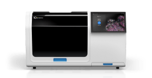
Xenium In Situ Analyzer
Simple design and interface plus fast run times and on-instrument analysis combine for world-class in situ profiling.
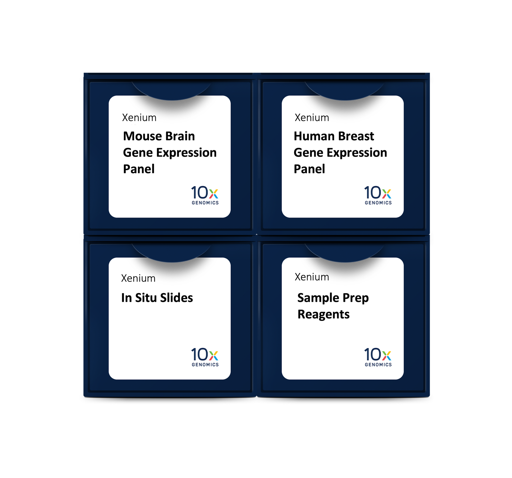
Xenium In Situ reagents and panels
Choose from a diverse menu of curated panels, customizable to fit your research needs.
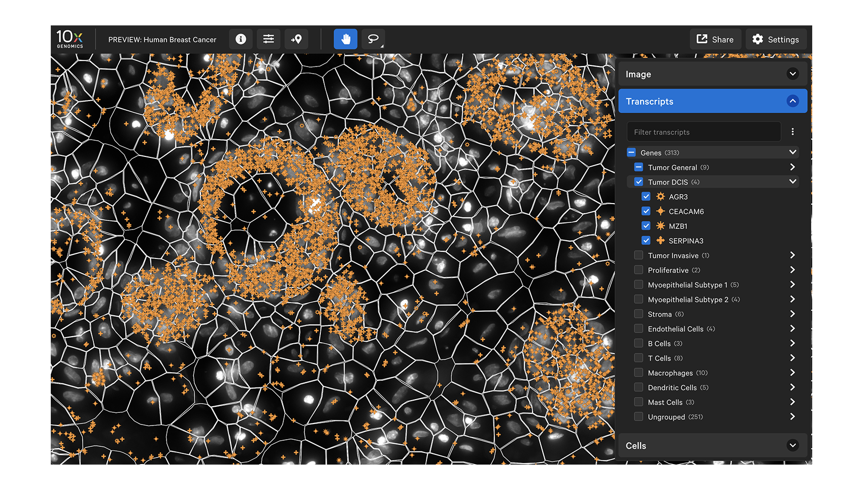
Data visualization
Xenium Explorer provides off-instrument interactive visualization, simplified data QC, and exploration of subcellular readouts.

Dedicated team of spatial experts
Get the help you need from our expert support team, reachable by phone or email.
